Biomedical Engineering Blog

Biomedical Engineering

The Biomedical Engineering blog is the place for conversation and discussion about topics related to engineering principles of the medical field. Here, you'll find everything from discussions about emerging medical technologies to advances in medical research. The blog's owner, Chelsey H, is a graduate of Rensselaer Polytechnic Institute (RPI) with a degree in Biomedical Engineering.

Previous in Blog: Ten Apples a Day Keeps the Doctor Away   Next in Blog: Documenting the Smell of History
Close
Close
Close
20 comments

New Tool to Diagnose Skin Cancer

Posted April 12, 2017 10:53 AM by Chelsey H

As summer approaches, it’s time to start stocking up on sunscreen. Skin cancer is the most common, but highly treatable, cancer, especially if detected early.

For everyone concerned with his or her own risk of skin cancer, there will soon be a quick, non-invasive test for diagnosis. Rather than taking a biopsy, which must be examined by a pathologist, the new system involves simply looking through a multiphoton microscope at the patient’s skin and determining whether it is cancerous or not.

An international group of scientists and engineers did the research and reported their findings in the November 2016 issue of Science Translational Medicine. The team found that mitochondria behave very differently in healthy versus cancerous tissue.

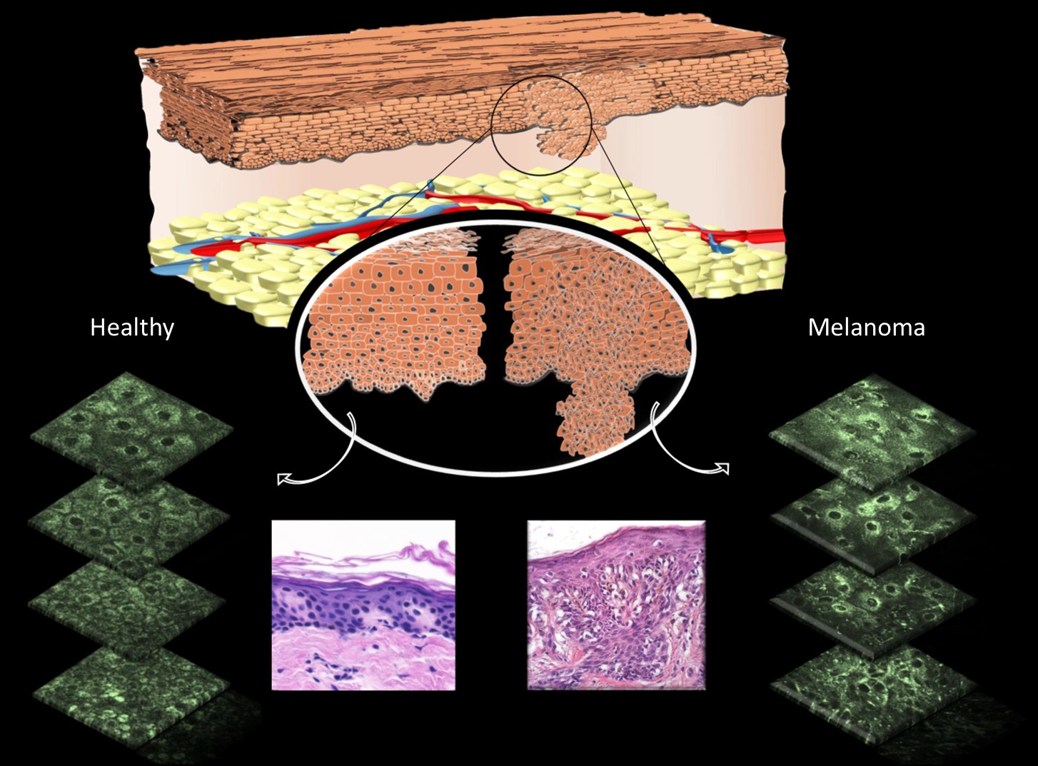
Mitochondria, small organelles that produce energy in cells, use a molecule called nicotinamide adenine dinucleotide (NADH) to produce energy for the cell. Because NADH naturally fluoresces without injecting any dye or contract, it can be observed using multiphoton microscopy to provide diagnostically useful information about the organization of the mitochondria in skin cells.

Dr. Irene Georgakoudi, senior author on the paper, noted that the multiphoton microscope uses lasers to obtain very high-resolution images of individual cells without having to slice the tissue physically. Normal cells in the mitochondria spread throughout the cell in a web-like pattern. Mitochondria in cancerous skin cells form clumps or clusters typically at the center of the cell along the border of the nucleus.

The small study tested 10 patients with skin cancer and four who did not have skin cancer. The imaging technique results were compared to the traditional biopsy results. The results demonstrated that the imaging technique correctly identified skin cancer in all 10 cancer patients, and made no false diagnoses in the four individuals without skin cancer.

Georgakoudi is hopeful that the test could be routinely used in doctor’s offices within five years as long as the price tag for the laser used in the microscope decreases. "Less-expensive lasers are on the horizon," concludes Georgakoudi. "However, this approach would enable a doctor to make a quick diagnosis and begin treatment immediately, which could ultimately lower health care costs associated with these very common cancers."

Materials provided by National Institute of Biomedical Imaging and Bioengineering.

Reply

Interested in this topic? By joining CR4 you can "subscribe" to
this discussion and receive notification when new comments are added.

Comments rated to be "almost" Good Answers:

Check out these comments that don't yet have enough votes to be "official" good answers and, if you agree with them, rate them!
Guru
Safety - Hazmat - New Member United States - US - Statue of Liberty - New Member Engineering Fields - Chemical Engineering - Old Hand

Join Date: Mar 2011
Location: Lubbock, Texas
Posts: 14331
Good Answers: 162
#1

Re: New Tool to Diagnosis Skin Cancer

04/12/2017 11:33 AM

Sounds like they need to really get on reducing the price of those lasers! This is really a great diagnostic breakthrough. Thanks for sharing.

__________________
If it ain't broke, don't fix it. Just build a better one.
Reply
Guru

Join Date: Jun 2009
Location: South of Minot North Dakota
Posts: 8378
Good Answers: 775
#2
In reply to #1

Re: New Tool to Diagnosis Skin Cancer

04/12/2017 2:17 PM

I once cut myself rather bad and thought I might need stitches.

I checked with WebMD and according to them I had cancer.

I hope the new laser tech here is a bit more accurate.

Reply
Guru
Hobbies - CNC - New Member Hobbies - DIY Welding - New Member Engineering Fields - Electromechanical Engineering - New Member

Join Date: Aug 2007
Posts: 23502
Good Answers: 419
#3
In reply to #2

Re: New Tool to Diagnosis Skin Cancer

04/12/2017 2:56 PM

so,... how's your home built chemo remedy coming along for your cancer...

__________________
“ When people get what they want, they are often surprised when they get what they deserve " - James Wood
Reply
Guru

Join Date: Jun 2009
Location: South of Minot North Dakota
Posts: 8378
Good Answers: 775
#4
In reply to #3

Re: New Tool to Diagnosis Skin Cancer

04/12/2017 3:12 PM

Once the main bleeding stopped some superglue and bandaids did the trick!

Cancer free so far! (no scar either!)

Reply
Guru
Hobbies - CNC - New Member Hobbies - DIY Welding - New Member Engineering Fields - Electromechanical Engineering - New Member

Join Date: Aug 2007
Posts: 23502
Good Answers: 419
#5
In reply to #4

Re: New Tool to Diagnosis Skin Cancer

04/12/2017 3:49 PM

glad to hear no duct tape was needed....

__________________
“ When people get what they want, they are often surprised when they get what they deserve " - James Wood
Reply
Guru

Join Date: Dec 2016
Posts: 2914
Good Answers: 115
#7
In reply to #4

Re: New Tool to Diagnosis Skin Cancer

04/12/2017 6:08 PM

And remember, if all else fails you can always cauterise it with a burning tyre.

Reply Score 1 for Good Answer
Guru

Join Date: Dec 2016
Posts: 2914
Good Answers: 115
#6
In reply to #1

Re: New Tool to Diagnosis Skin Cancer

04/12/2017 5:47 PM

NADH absorbs light of wavelength 340±30 nm and fluoresces (pdf) at 460±50 nm, and so they're using a UV laser. There are comparatively inexpensive diode lasers that emit in this range (360 nm, 1 to 50 mW) and they're paying $100K for this thing? WTH for?

"Less-expensive lasers are on the horizon," concludes Georgakoudi.

They're not 'on the horizon', they're here right now and practically sitting in his lap.

Reply
Guru

Join Date: Mar 2007
Location: by the beach in Florida
Posts: 33231
Good Answers: 1807
#8
In reply to #6

Re: New Tool to Diagnosis Skin Cancer

04/12/2017 9:14 PM

Well that looks like pretty high magnification, that and what is the intensity of light, might be a tricky combination for viewing at high speed scanning level that would be practical....and mapping the spot and area on the skin that is suspected should be automated....there might be a lot more to this then just a laser and microscope...when they say "cheaper lasers" they probably are speaking about the scanning machine itself, the whole machine...

__________________
All living things seek to control their own destiny....this is the purpose of life
Reply
Guru

Join Date: Dec 2016
Posts: 2914
Good Answers: 115
#9
In reply to #8

Re: New Tool to Diagnosis Skin Cancer

04/12/2017 9:54 PM

Controlling the intensity of the light is trivial. You start out with more light than you need and insert a variable-density UV filter in the optical path, split the beam into two parts post-filter, supplying one part to a photodiode which is part of servo loop that controls the filter density (using by rotating it), thus maintaining the intensity at the setpoint. You can also control the diode current directly, but other effects such beam coherence and, especially troublesome, wavelength-shift, come into play, complicating control. Filtering the beam is straightforward and highly predictable.

As far the equipment goes, yes, multiphoton microscopes are expensive, but they cited 'laser' specifically and several times. I suspect they're currently using a nitrogen or similar UV gas laser, which are quite expensive.

Reply
Guru

Join Date: Dec 2016
Posts: 2914
Good Answers: 115
#11
In reply to #10

Re: New Tool to Diagnosis Skin Cancer

04/13/2017 1:27 AM

Great links. Thanks.

Reply
Guru
Safety - Hazmat - New Member United States - US - Statue of Liberty - New Member Engineering Fields - Chemical Engineering - Old Hand

Join Date: Mar 2011
Location: Lubbock, Texas
Posts: 14331
Good Answers: 162
#14
In reply to #10

Re: New Tool to Diagnosis Skin Cancer

04/13/2017 9:25 AM

It seems clearer to me right now, that in situ measurement may indeed require femtosecond time resolution. Perhaps we are still talking Ar+ laser, N2 laser pumped dye laser with Q switching, etc.?

Recently, there were announcements of advances in LED or other solid-state lasers, that warrant checking into with respect to advanced biophotonics.

__________________
If it ain't broke, don't fix it. Just build a better one.
Reply
Guru
Safety - Hazmat - New Member United States - US - Statue of Liberty - New Member Engineering Fields - Chemical Engineering - Old Hand

Join Date: Mar 2011
Location: Lubbock, Texas
Posts: 14331
Good Answers: 162
#13
In reply to #8

Re: New Tool to Diagnosis Skin Cancer

04/13/2017 9:19 AM

Actually, I am starting to wonder if it "scans" an area, or takes a spatial coherence snap-shot, and then the phase is transformed in a FFT (computer), to reveal spatial constructs. I don't even see how a patient can hold still long enough for the spatial resolution required, unless the lasers are TEM00 and are fast pulse. In fact, that might be the problem with the high cost of the lasers. LED lasers available might not be operating in spatial mode TEM00, or have sufficiently fast pulsing to make this work, but I already know there are mode switches and Q switches, etc, heck, even mechanical shutters, or dichroic shutters that could make this work.

__________________
If it ain't broke, don't fix it. Just build a better one.
Reply
Guru
Safety - Hazmat - New Member United States - US - Statue of Liberty - New Member Engineering Fields - Chemical Engineering - Old Hand

Join Date: Mar 2011
Location: Lubbock, Texas
Posts: 14331
Good Answers: 162
#12
In reply to #6

Re: New Tool to Diagnosis Skin Cancer

04/13/2017 9:13 AM

Did you notice in the article they were doing high spatial resolution imaging, and using two photon spectroscopy? That probably means the wavelength employed is nearer 680 nm, but they should still observe the 460 nm band fluoresce.

I agree it is total BS that laser should or does cost what they say it does. Another example of medical research where money and common sense are no object.

I would say those lasers they think they are waiting for do not have to be imported from the King of Knossos for them to work, gold plated with platinum overlay or not.

Maybe Gerogakoudi is just trying to make money for Greece.

__________________
If it ain't broke, don't fix it. Just build a better one.
Reply
Guru

Join Date: Mar 2007
Location: by the beach in Florida
Posts: 33231
Good Answers: 1807
#15

Re: New Tool to Diagnose Skin Cancer

04/13/2017 10:56 AM

"Traditionally, confocal and multiphoton-excited fluorescence (MPEF) microscopy images are formed by scanning a near-diffraction-limited, micron-size laser spot over a volume of interest that measures hundreds of microns. The latest multiphoton microscopes are equipped with fast galvanometer or resonant scanners able to generate hundreds of frames per second. The information in each voxel is acquired by accumulating the fluorescence resulting from the nonlinear absorption of many femtosecond (fs) laser pulses in the near-infrared (NIR) wavelength range, typically 900–1300 nm. To ensure that a sufficient number of consecutive laser pulses contribute to each voxel, the ultrafast source should have a relatively high (greater than 20 MHz) pulse repetition rate (PRR).

Conversely, the practical driver for fluorescence intensity, image brightness, and contrast in MPEF microscopy is the laser peak power delivered to the sample. Laser peak power is a measure of instantaneous photon density:

For most bioimaging applications—especially in vivo imaging tasks—researchers limit laser average power to hundreds of milliwatts (mW) at most to prevent specimen damage through heating and/or phototoxicity. Peak power (and consequently the fluorescence intensity) can be increased by decreasing either the PRR or the pulsewidth. For practical purposes, nearly all laser scanning multiphoton microscopes today are equipped with ultrafast lasers operating at a PRR between 50 and 100 MHz and peak powers on the order of hundreds of kilowatts—the sweet spot for today's point-scanning MPEF imaging applications."

"In recent years, three-photon excitation fluorescence (3PEF) microscopy has been proposed as a promising imaging modality to increase penetration depth in vivo, especially in neuroscience for the functional imaging of subcortical structures.5 The three-photon absorption process dictates that the laser light be shifted further into the NIR (typically to 1.3 μm and in some cases up to 1.7 μm), enabling greater reduction of scattering effects and allowing ballistic photons to make their way through the live tissue to the imaging plane.

The nonlinear three-photon process is a very low-probability quantum event that requires the simultaneous presence and absorption of three NIR photons to generate a fluorescence signal. Researchers rely on high-peak-power ultrafast lasers to compensate for such low probability. Ytterbium-doped ultrafast amplifiers—equipped with an OPA or NOPA to shift the excitation to the adequate wavelength—offer a robust and dependable solution."

http://www.bioopticsworld.com/articles/print/volume-8/issue-8/biooptics-features/ultrafast-lasers-ultrahigh-peak-power-femtosecond-lasers-advance-bioimaging.html

__________________
All living things seek to control their own destiny....this is the purpose of life
Reply
Guru
Safety - Hazmat - New Member United States - US - Statue of Liberty - New Member Engineering Fields - Chemical Engineering - Old Hand

Join Date: Mar 2011
Location: Lubbock, Texas
Posts: 14331
Good Answers: 162
#16
In reply to #15

Re: New Tool to Diagnose Skin Cancer

04/13/2017 11:46 AM

Based on the limitations of peak power, it might appear what is needed is a better detector than a better cheaper laser. Did someone make an advance recently in single photon detection?

__________________
If it ain't broke, don't fix it. Just build a better one.
Reply
Guru

Join Date: Mar 2007
Location: by the beach in Florida
Posts: 33231
Good Answers: 1807
#17
In reply to #16

Re: New Tool to Diagnose Skin Cancer

04/13/2017 12:19 PM
__________________
All living things seek to control their own destiny....this is the purpose of life
Reply
Guru
Safety - Hazmat - New Member United States - US - Statue of Liberty - New Member Engineering Fields - Chemical Engineering - Old Hand

Join Date: Mar 2011
Location: Lubbock, Texas
Posts: 14331
Good Answers: 162
#18
In reply to #17

Re: New Tool to Diagnose Skin Cancer

04/13/2017 1:53 PM

Well, that is as near SPD as it needs to be, just in the wrong spectral band, I think.

I think they will easily overcome that minor hurdle if not already.

It used to be that a SPD system was actually only able to detect 10-20 photons based on catching one event with about 5-10% quantum efficiency.

They called it "single-photon" because the energy of the photoelectrons captured was consistent with that, and they could use event counting statistics.

__________________
If it ain't broke, don't fix it. Just build a better one.
Reply
Participant

Join Date: Apr 2017
Location: Singapore
Posts: 1
#19

Re: New Tool to Diagnose Skin Cancer

04/18/2017 10:32 AM

but some people says cancer is because our body is too acidic :-(

Reply
Guru
Safety - Hazmat - New Member United States - US - Statue of Liberty - New Member Engineering Fields - Chemical Engineering - Old Hand

Join Date: Mar 2011
Location: Lubbock, Texas
Posts: 14331
Good Answers: 162
#20
In reply to #19

Re: New Tool to Diagnose Skin Cancer

04/18/2017 11:29 AM

Too acidic, or not acidic enough? I think the human body regulates pH very effectively, unless there is a terribly bad metabolic disorder, such as ketoacidosis. Breathing too rapidly (due to pain, etc.) may result in breath alkalosis.

I know that never drinking beer gives me Beernemia, and too much beer results in Beernitis, Beeratosis, Beermyopia, and other conditions, related to not showing up at work on time, etc.

If too acidic, please do breathing excercises, or cut back slightly on potassium.

__________________
If it ain't broke, don't fix it. Just build a better one.
Reply
Reply to Blog Entry 20 comments
Interested in this topic? By joining CR4 you can "subscribe" to
this discussion and receive notification when new comments are added.

Comments rated to be "almost" Good Answers:

Check out these comments that don't yet have enough votes to be "official" good answers and, if you agree with them, rate them!
Copy to Clipboard

Users who posted comments:

aloha168888 (1); Andrew Westman (4); James Stewart (7); phoenix911 (2); SolarEagle (4); tcmtech (2)

Previous in Blog: Ten Apples a Day Keeps the Doctor Away   Next in Blog: Documenting the Smell of History

Advertisement